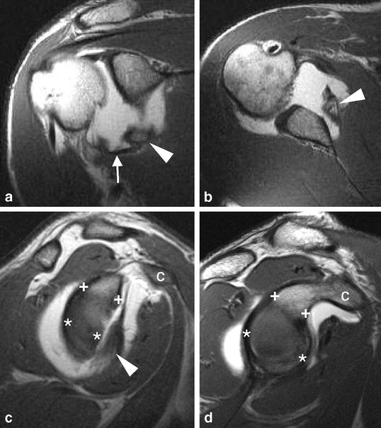
Fig. 4a–d.

Bony Bankart lesion. a Coronal oblique and b transverse T1-weighted MR arthrograms show avulsion of a large osseous fragment (arrowheads) from the glenoid together with the attaching anteroinferior labrum and IGHL (arrow). c Sagittal oblique T1-weighted MR arthrogram demonstrates “inverted peer” geometry of glenoid in presence of a “relevant” bony Bankart lesion (arrowhead). Note smaller anteroposterior diameter of glenoid below (**) midglenoid notch than above (++) it. For comparison, normal peer-shaped configuration of glenoid is shown on d sagittal oblique T1-weighted MR arthrogram from individual with stable shoulder (C coracoid process)
