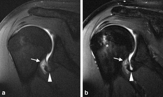
Fig. 8a, b.

HAGL lesion. a Coronal oblique fat-suppressed T1-weighted MR arthrogram and b corresponding intermediate weighted TSE image demonstrate avulsion of the IGHL at its humeral insertion causing a J-shaped configuration of the axillary recess (arrowheads). Contrast extravasation can be seen at the site of the tear (arrows). Bone marrow edema within humeral head (*) was caused by acute Hill-Sachs defect (not shown)
