
Fig. 1a, b.
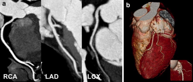
Dual-source CT coronary angiography in a 62-year-old woman with suspected coronary artery disease (mean heart rate during scanning 76 bpm, Agatston score 0). a Curved thin-slab maximum-intensity projections through the centerline of the right coronary (RCA), left anterior descending (LAD), and left circumflex artery (LCX). Slight blurring of the mid-RCA and mid-LAD rendered image quality as good (score 2) in these segments, while image quality was rated excellent (score 1) in all other segments. Coronary artery disease could be reliably excluded in this patient. b Volume-rendered image of the left coronary arteries and of the proximal RCA (insert) demonstrates accurate depiction of the coronary artery tree