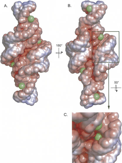
FIGURE 8.
Electrostatic surface potential of the lowest-energy NMR structure with associated Mn2+. Electrostatic potential of the RNA displayed on the solvent accessible surface with the following scale: red = −9 kT/e, white = −2 kT/e, and blue = 5 kT/e. Manganese ions are shown as green spheres with the radius of a hexahydrated ion. (B) Rotation (180°) of A. (C) Detailed view of the RNA tilted 50° on the x-axis to display the Mn2+ ion bond in the middle of the helix (not visible in other views).

