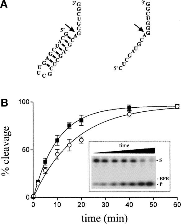
FIGURE 6.
Cleavage assays of differently structured substrates. (A) Secondary structures and nucleotide sequences of the substrate including a 5′-end hairpin, as well as that of its single-stranded counterpart. The arrows indicate the cleavage sites. (B) Graphical representation of the cleavage percentages as a function of time for both substrates, the single-stranded substrate (closed squares) and the substrate with the 5′-end hairpin (open circles). The inset shows a typical autoradiogram of a denaturing 20% PAGE gel for the cleavage of the substrate with the 5′-end hairpin. The positions of the xylene cyanol (XC), the substrate (S), and the product (P) are indicated.

