Fig.2.

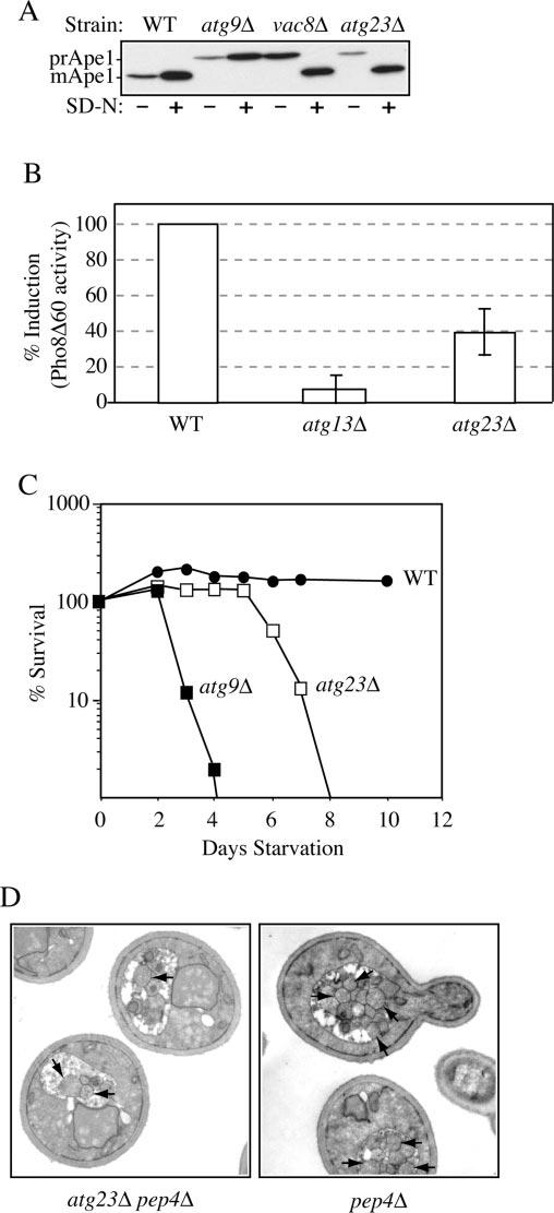
Atg23 is required for autophagy. A, precursor Ape1 matures in atg23Δ cells during starvation conditions. Wild type (WT) (SEY6210), atg23Δ (KTY14), atg9Δ (JKY007), and vac8Δ (D3Y102) cells were grown to A600 = 1.0 in SMD and then kept in SMD or starved in SD-N medium for 2 h. Protein extracts were prepared and subjected to immunoblot analysis with anti-Ape1 antiserum. B, autophagy is partially induced in atg23Δ cells. Wild type (TN124), atg23Δ (KTY9), and atg13Δ (D3Y103) cells were shifted from SMD to SD-N medium for 4 h. Autophagy was measured by the levels of Pho8Δ60 activity in whole cell protein extracts. Activity in the wild type strain was set to 100% and activity in the other strains normalized relative to wild type. Error bars represent the S.D. from three separate experiments. C, the atg23Δ cells exhibit intermediate starvation resistance. Wild type (SEY6210), atg23Δ (KTY14), and atg9Δ (JKY007) cells were grown in SMD to A600 = 1.0 and then shifted to SD-N. At the indicated day, viability was determined by removing aliquots, plating in triplicate, and counting the number of colonies per plate after 2–3 days growth. D, fewer autophagic bodies accumulate in atg23Δ pep4Δ cells during starvation conditions. Cells from the pep4Δ (TVY1) and pep4Δ atg23Δ (KTY22) strains were grown in SMD, then shifted to SD-N for 4 h, fixed in potassium permanganate, and processed for electron microscopy as described under “Experimental Procedures.” Arrows indicate autophagic bodies.
