Fig .7.

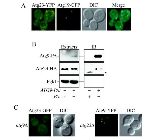
Atg23-GFP and Atg9-YFP exhibit similar localization as well as physical and functional interactions. A, Atg9 and Atg23 localize to several punctate structures dispersed in the cytoplasm. Cells containing Atg9-YFP and CFP-Atg8 integrated at their chromosomal loci (FRY134) or those containing both Atg23-YFP integrated at the chromosomal ATG23 locus (KTY26) and the plasmid bearing Atg19-CFP were grown in selective medium to A600 = 0.8–1.2 and imaged with a fluorescent microscope. B, Atg23 interacts with Atg9. Atg23-HA (KTY8) cells transformed with Atg23-HA and either Atg9-PA (KTY74) or pRS416-CuProtA as a control were used to prepare detergent-solubilized extracts as described under “Experimental Procedures.” IgG-Sepharose beads were used to affinity-purify the PA fusions together with the associated proteins. Eluted polypeptides were separated by SDS-PAGE and then visualized by immunoblotting (IB) with antiserum to PA, HA, and Pgk1. For each experiment, 0.013% of the total lysate or 4.45% of the total eluate was loaded per gel lane. The asterisk marks a contaminating band. C, Atg9 is required to recruit Atg23 to membranes. The atg9Δ mutant bearing an integrated Atg23-GFP (KTY32) or Atg9-YFP cells deleted for the ATG23 ORF (KTY51) were grown and imaged as in A. DIC, differential interference contrast.
