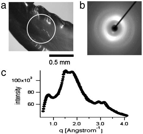
Fig. 2.
XRD on Nereis jaw. (a) Light microscopy image of the sample as mounted in the XRD instrument. The white circle denotes the diameter of the x-ray beam. (b) Typical XRD pattern of Nereis jaw. The diffraction signal is anisotropic with a preferred orientation along the axis of the jaw. (c) Integration of the two-dimensional diffraction pattern in b over the azimuth yields a curve of intensity versus the scattering vector q. Intensity is in arbitrary units.

