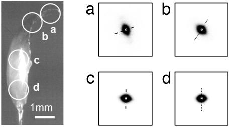
Fig. 3.
SAXS on Nereis jaw. (Left) Light microscopy image of a jaw mounted in the SAXS apparatus. The white circles indicate the approximate positions and diameter of sampled areas. (Right a–d) SAXS patterns obtained from each point circled in Left. The anisotropic signal is indicative of a fiber-like structure, where the longitudinal axis of the fibers is oriented along the short axis of the scattering pattern (dashed line). The fiber axis follows the outline of the jaw.

