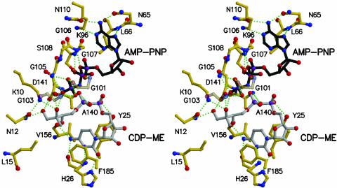
Fig. 4.
Stereoview of the active site. Bonds in AMP-PNP are black, bonds in the protein are yellow, and bonds in CDP-ME are gray. Selected hydrogen-bonding interactions are shown as green dashed lines. Water molecules have been omitted; Gly-101 and Gly-103 are semitransparent for the purpose of clarity.

