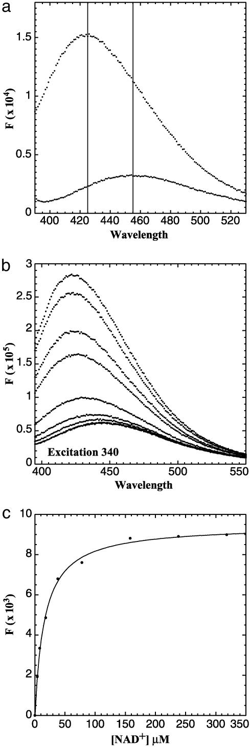
Fig. 5.
(a) Blue shift and enhanced fluorescence of bound NADH. Free NADH has a peak emission at 455 nm when excited at 340 nm (lower trace). Addition of CtBP results in a shift of the emission peak to 425 nm and an increase in the quantum yield (upper trace). (b) NAD+ displacement of NADH. The emission scan of CtBP bound with NADH excited at 340 nm (top trace). The titration of NAD+ resulted in a loss of NADH fluorescence. At saturating levels of NAD+, an emission scan typical of free NADH was observed. (c) Plot of NAD+ versus ΔF. Data were fit with Eq. 2. See Materials and Methods for calculation of Kd.

