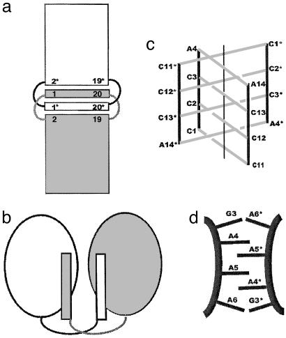
Fig. 1.
Intercalation and domain swapping in nucleic acids and proteins. (a) Schematic diagram of this intercalated RNA·DNA structure. Helices stack head to head. Base pair 1*·20* of one helix intercalates between base pairs 1·20 and 2·19 of the other. Similarly, base pair 1·20 intercalates between base pairs 1*·20* and 2*·19* (* indicates symmetry-related molecule). (b) Schematic of 3D domain swapping as seen in proteins. (c) The central I-DNA motif region of the crystal structure of d(CCCAAT). Two strands are in the asymmetric unit, and an additional duplex (marked with asterisks) completes the four-stranded intercalated motif of the molecule. C·C base pairing is extended to A·A base pairs in this intercalation. (d) The schematic diagram of the zipper-like motif found in the middle of the crystal structure of d(GCGAAAGCT) with alternate intercalations of unpaired adenines and doubling of the interspacing found in this region.

