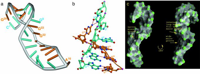
Fig. 3.
Structure of the RNA aptamer. (a) Overall structure of the α-p50 RNA aptamer. Residues 1–15 representing one strand are cyan, and residues 16–29 representing the other strand are orange. (b) A close-up view of the folded structure of the internal loop. Three cross-strand-stacked guanines are indicated. The non-Watson–Crick hydrogen bond pairing between U6 and C24 and between A9 and G22 of the internal loop are indicated. (c) Surface presentation (23) of the RNA in two orientations related by 180° rotation around the long axis. The image on the left is in the same orientation as a and shows the narrow minor-groove section near the internal loop. The image on the right shows the protein-binding groove of the RNA.

