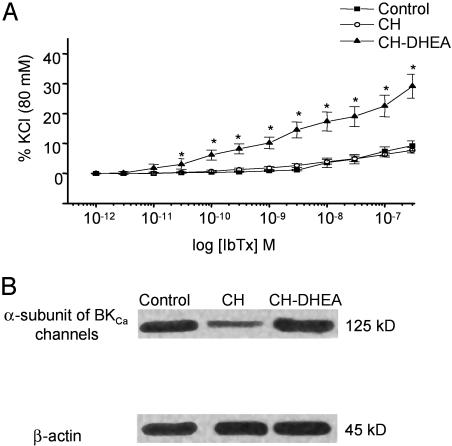
Fig. 7.
Effects of DHEA on PA reactivity to BKCa blockers and BKCa expression. (A) In vitro concentration–response curves for the effect of IbTx on the resting tension of IPA rings from CH and CH-DHEA rats. The amplitude of contraction is expressed as a percentage of the KCl (80 mM)-induced response obtained at the beginning of the experiments. Note the increase in the IbTx response in rings from CH-DHEA rats. Data points are mean ± SEM for CH (n = 11, N = 4) and CH-DHEA (n = 11, N = 4) rats. (B) Immunoblots of the BKCa α-subunit after 21 day of oral administration of DHEA. Each lane was loaded with 10 μg of protein. BKCa α-subunit was recognized by the Ab as a 125-kDa immunoreactive band. BKCa α-subunit is down-regulated in CH vs. control groups, whereas its expression was similar between control and CH-DHEA groups. No difference was observed in the 45-kDa β-actin bands used as an internal standard.

