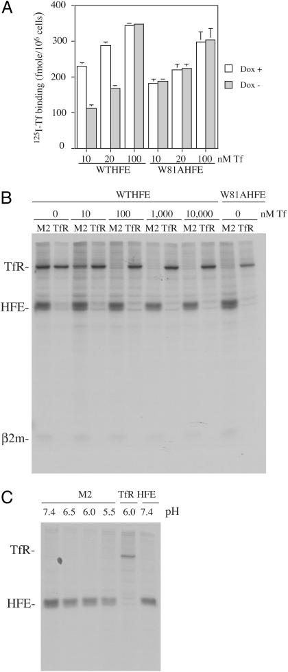
Fig. 1.
Evidence for the competition between Tf and HFE for binding to the TfR in intact cells and cell lysates. (A) Binding of 125I-Tf to cells. fWTHFE/tTA and fW81AHFE/tTA HeLa cells expressing HFE (Dox-) or not (Dox+) were incubated with 10, 20, and 100 nM 125I-Tf for 90 min at 4°C to allow binding to cell surface TfRs. Excess unlabeled diferric Tf (200×) was added to measure nonspecific binding. Samples were done in triplicate. (B) Coimmunoprecipitation of HFE in the presence of Tf. fWTHFE/tTA HeLa cells expressing HFE (Dox-) were labeled overnight with 50 μCi/ml [35S]methionine/cysteine (1 Ci = 37 GBq). Cell lysates were preabsorbed with protein A beads, and diferric Tf was added for 30 min at 4°C, followed by immunoprecipitation with an anti-Flag antibody (M2) for HFE or a mouse monoclonal TfR antibody. The experiment was repeated twice with similar results. (C) Coimmunoprecipitation of fW81AHFE at pH 7.4, 6.5, 6.0, and 5.5. fW81AHFE/tTA cells (Dox-) were labeled with [35S]methionine/cysteine as described above. For each sample, pH was kept the same throughout the entire immunoprecipitation and washes. The same antibodies were used as above except in the last lane, where a mouse monoclonal (8C10) against HFE was used as a control.

