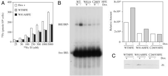
Fig. 3.
fW81AHFE/tTA HeLa cells have a low iron phenotype. (A) Tf-55Fe accumulation in cells. Tf-55Fe was added to the complete culture medium of cells expressing (Dox-) or not expressing (Dox+) HFE to the final concentrations of 25, 50, 100, 250, 500, 1,000, and 5,000 nM. Cells were incubated for 20 h at 37°C at 5% CO2. They were solubilized after acid wash at 4°C to strip off surface-bound Tf. The internal levels of 55Fe were expressed as pmol per 106 cells. Because of the similar uptake rates between fW81AfHFE/tTA and fWTHFE/tTA HeLa (Dox+), the results were averaged together and labeled Dox+. The experiment was repeated with similar results. (B) Gel-shift analysis of IRP binding to IREs by fWTHFE/tTA (WT), fW81AHFE/tTA (W81A), and fC260YHFE/tTA (C260Y) HeLa cell extracts. Extracts from cells expressing (Dox-) or not expressing (Dox+) HFE were incubated with 32P-labeled IRE as described in Experimental Methods and subjected to PAGE. Quantitative analysis of the IRE/IRP complexes in B was done by using a PhosphorImager. (C) Western blot of Ft levels using the same cell extracts as for IRP binding analysis in B. Thirty micrograms of lysate protein was loaded onto each lane. Blots were probed with an antibody to Ft as described in Experimental Methods. The experiments shown in B and C were repeated with similar results.

