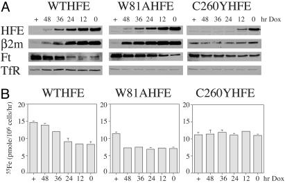
Fig. 5.
The effect of the level of HFE expression on Tf-mediated iron uptake into cells. (A) Western blots of HFE, β2m, Ft, and TfR. Cell lines expressing HFE were treated for 0–48 h or maintained continuously with Dox (1 μg/ml) to turn off HFE expression. At each time point they were solubilized and subjected to Western analysis with the indicated antibodies. For all of the Western blots, 30 μg of protein was loaded into each lane. (B) Tf-55Fe uptake. Tf-55Fe uptake was measured after cells were incubated with Dox to turn off HFE expression for the same time points as in A. 55Fe-Tf (100 nM) was used. The results of the 1-h incubation at 37°C are presented for simplicity, although linear uptake was always observed for at least 3 h. The rates of uptake are expressed as pmol 55Fe per 106 cells per h. Nonspecific background was performed the same as above except that the incubation was performed on ice. Samples were always in triplicate and the experiment was repeated with similar results.

