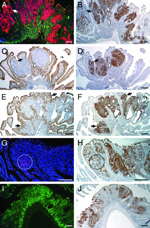
Fig. 2.
Analysis of clusterin expression patterns. (A) Dual-staining immunofluorescence assay on a Min tumor section demonstrated that the WT Apc antigen (green) was present in the normal crypts, but not in tumor cells, where a high level of β-catenin antigen (red) was detected. (B) Staining of clusterin RNA (dark brown) with antisense probe in an adjacent section is similar to that of cytoplasmic β-catenin protein. The arrows in A and B point to a small exceptional region lacking both WT Apc antigen and clusterin antigen. (C–F) Expression of differentiation markers and clusterin RNA in intestinal tumors. (A, C, E, G, and I) RNA signals (dark brown) of two differentiation markers, intestinal alkaline phosphatase (C) and small proline-rich protein 2a (E), with their antisense probes. (B, D, F, H, and J) Sections adjacent to C and E were hybridized for clusterin RNA (dark brown) with the antisense probe for comparison (D and F). Solid arrows in C–F point to small exceptional regions expressing one of the differentiation markers and clusterin, whereas open arrows point to small regions lacking both of them. (G) Cells undergoing apoptosis within tumors were labeled red by fluorescent TUNEL assay, in which the nuclei were stained blue by 4′,6-diamidino-2-phenylindole (DAPI). (H) For comparison, a section adjacent to G was hybridized for clusterin RNA (dark brown) with the antisense probe. Circles indicate the regions showing a high apoptotic index, where clusterin RNA levels were relatively low. (I) A section of tumor stained for Ki67 antigen by immunofluorescence (green signal). (J) A section adjacent to I hybridized for clusterin RNA (dark brown) with the antisense probe. (Bars: 200 μm.)

