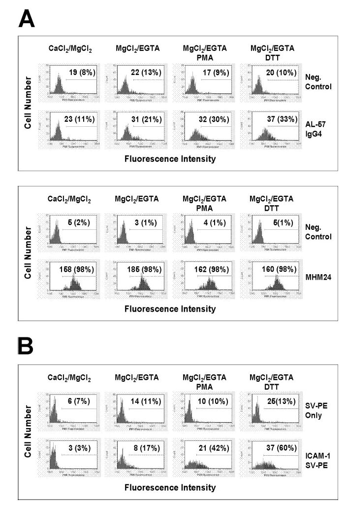
Fig. 3.

AL-57 IgG4 and ICAM-1 bind to the activated HA form of LFA-1 on PBMCs. Binding of AL-57 IgG4 (A) and ICAM-1 (B) to PBMCs with inactivating buffer (CaCl2/MgCl2) or with activating buffer (MgCl2/EGTA) in the presence or absence of PMA (10 ng/ml) or DTT (500 μM) was determined by flow cytometric analysis as described in Materials and Methods. The concentration of each IgG or ICAM-1 used was 10 μg/ml. For AL-57 staining, Neg. Control indicates negative control with just the secondary PE-labeled antibody used for staining; MHM24 was used as a control that did not distinguish between the HA and LA forms of LFA-1. For ICAM-1 staining, a multimeric complex [ICAM-1/streptavidin (SV)-PE] of biotinylated human ICAM-1-Fc and PE-labeled streptavidin was used; SV-PE only indicates negative control with just the PE-labeled streptavidin used for staining. Shown here are representative histograms of samples under indicated conditions. The x-axis depicts the fluorescence intensity of individual cells, and the y-axis represents the cell number. The numbers shown are relative values of MFI with relative percentages of positive cells in parenthesis.
