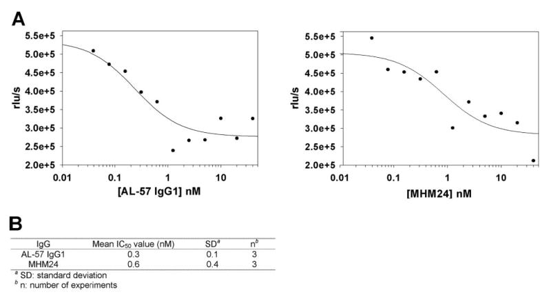
Fig. 7.

AL-57 IgG1 inhibits PHA-induced cell proliferation. PBMCs were treated with PHA at 1 μg/ml in the presence of Mg2+, serially diluted IgG for 3 days, and then analyzed for proliferation using a BrdU chemiluminescence assay. (A) IC50 determination of AL-57 IgG1 and MHM24. Relative luminescence units per second (rlu/s) from each sample were plotted as a function of the IgG concentration. From the representative plots shown here, IC50 values were calculated to be 0.2 nM for AL-57 IgG1 and 0.8 nM for MHM24, using SigmaPlot 8.0 software. (B) Summary of IC50 values from three independent experiments.
