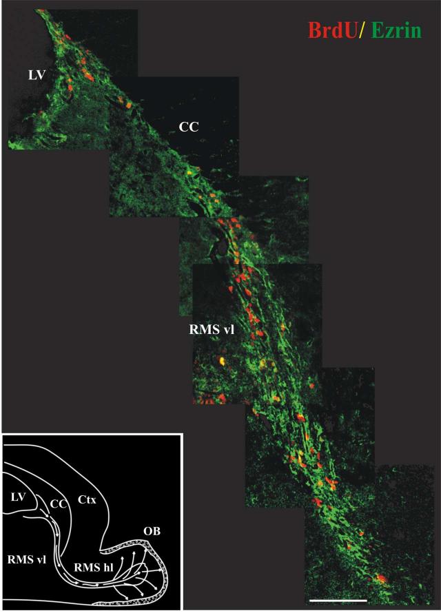
Figure 2.
Ezrin is expressed along the path of migrating cells in the RMS.
Montage of images starting from the SVZ and following the vertical limb of the RMS in a single thirty-micron sagittal section from an adult mouse sacrificed two hours after injection with BrdU. Sections were stained with antibodies to ezrin (green) and BrdU (red) and visualized by confocal microscopy after staining with fluorescently labeled secondary antibodies. As seen here, ezrin-immunopositive processes form a dense trabecular network surrounding the columns of BrdU-labeled migrating neuroblasts. These ezrin-immunopositive processes surround, but in most cases did not co-localize with, the BrdU-labeled neuroblasts. Inset: a schematic of the mouse forebrain illustrating the migratory pathway of SVZa derived neuroblasts en route to the olfactory bulb (adapted from (Coskun and Luskin, 2002). LV-Lateral Ventricle, CC- Corpus Callosum, Ctx - Cerebral Cortex, RMS vl - Vertical Limb of Rostral Migratory Stream, RMS hl - Horizontal Limb of Rostral Migratory Stream, OB - Olfactory Bulb. Scale bar = 100 μm.

