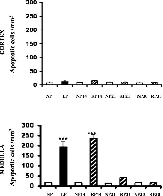
Fig 3.
Quantitation of apoptotic cells in medullary collecting tubules from kidneys of NP, LP, and RP fed rats. Ten consecutive fields were randomly selected in each renal cortex and medulla and were evaluated at 400× magnification. The number of apoptotic cells was expressed as apoptotic cells per square millimeter. Upper panel, Nonsignificant differences were observed in LP vs NP from kidney cortexes. Lower panel, Apoptosis peaked in cells from medullary collecting duct segments (OMCDs and IMCDs) in low-protein–fed rats for 14 days compared to that of NP, ***P < 0.001, n = 6. Persistent apoptotic nuclei in epithelial cells from medulla duct segments were shown after readministration of 24%protein in diet for 14 days. No significant increase of the apoptotic cell number was observed in medullary collecting ducts from kidneys after 21 and 30 days of protein recovery (RP)

