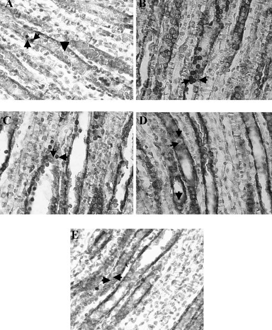
Fig 4.
Histologic sections of kidney medulla from normal protein (NP), low protein (LP), and recovery protein (RP) fed rats. Through colocalization staining of apoptotic nuclei (TUNEL technique) with HSP70 expression, apoptotic nuclei appeared as intense brown-staining nuclei and cytoplasmic slight brown HSP70 expression in the same tubule epithelial cells from medullary collecting ducts. Light counterstaining with 0.5% with hematoxylin was performed to reveal nonapoptotic nuclei, nuclei appeared as blue staining. (A) NP: Apoptotic cells were rarely seen in OMCDs and IMCDs (left pointing arrow). Slight Hsp70 staining was shown (up arrow), nonapoptotic nuclei appeared as blue staining (down arrow). (B) LP: Increased number of apoptotic nuclei, as intense brown-staining nuclei (right pointing arrow), and weak HSP70 cytoplasm expression were shown in medullary collecting ducts (left pointing arrow). (C) RP after 14 days: Persistently increased number of apoptotic epithelial cells in OMCDs and IMCDs (left pointing arrows), in addition to weak Hsp70 immunoreaction in the cytoplasm of the same epithelial cells (down arrow), were shown. (D) RP after 21 days: Low number of positive TUNEL cells were shown in epithelial cells of medullary collecting ducts (right arrow) with strong Hsp70 immunoreaction in the same tubular cells (upper downward arrow), nonapoptotic nuclei appeared as blue staining (lower downward arrow). (E) RP after 30 days: Apoptosis was absent in epithelial cells from medullary duct segments, after 30 days with weak Hsp70 immunoreaction expression similar to the one of NP (left pointing arrow). Nonapoptotic nuclei appeared as blue staining (right pointing arrow). Magnification, 630×. The colors mentioned in the text refer to the original specimens

