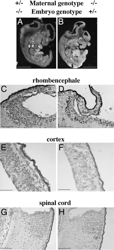
Fig. 2.
Longitudinal section of whole tph1 embryos 2 and 3 of Fig. 1. (A and B) Embryos 2 and 3 were obtained, respectively, from a cross between a tph1+/− mother and a tph1+/− father and a cross between a tph1−/− mother and a wild-type father. Microscopic investigations revealed flattening of the head region at the level of the IV ventricle in the embryos obtained from a null mother. The corresponding CNS area in an embryo obtained from a heterozygous mother showed a normal histology. (C and D) A 2-h BrdU pulse revealed a reduced number (30% less) of labeled cells in the ventricular zone of the heterozygous embryo obtained from a null mother as compared with a null embryo obtained from a heterozygous mother. The analysis also revealed a 24% reduction in BrdU labeling in the roof of neopallial cortex of heterozygous embryos from null mothers (compare F with E). No major difference in BrdU labeling was observed at the level of the spinal cord (G and H). (Scale bars: 0.25 cm in A and B and 100 μm in C–H.)

