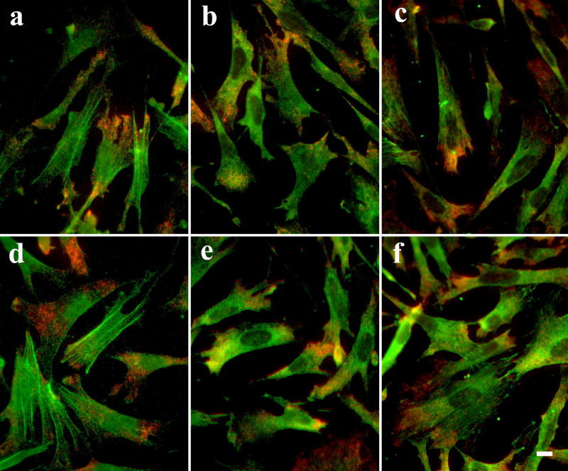
Fig. 8. No detectable deleterious effects on cell morphology and antigenicity in CEFs treated with HCl.
After detection of beta-actin mRNA (red), the cells were treated with either PBS (a-c) or 0.02 N HCl for 20 min before processed for immunostaining (green) for beta-actin (a, d), eEF-1A (b, e) or vinculin (c, f). Scale bar = 10 μm.

