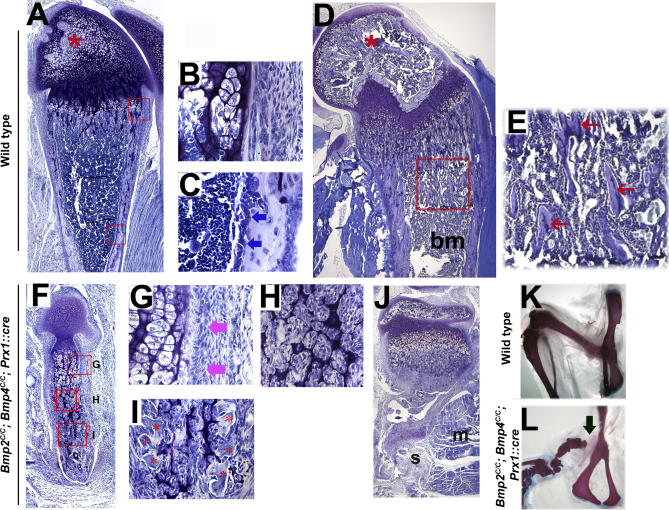
Figure 6. Defects in Bone Formation in the Absence of BMP2 and BMP4.
Toluidine blue staining of sagittal sections of distal femurs.
(A–E) Control femurs at 1 wk (A) and 3 wks (D) of age. Boxed areas in (A) are enlarged in (B) and (C). Boxed area in (D) is enlarged in (E). Blue arrows in (C) point to osteoblast cells lining the surface of cortical bone. Pink arrows in (G) show similar cells to those in (C) in Bmp2C/C; Bmp4C/C; Prx1::cre femurs. Red star in (A) and (D) marks the secondary ossification center. Pale blue–stained tissue in (E), marked by red arrows, is trabecular bone. bm, marks the bone marrow cavity in (D).
(F–J) Bmp2C/C; Bmp4C/C; Prx1::cre femurs at 1 wk (F) and 3 wks (J) of age. Boxed areas in (F) are enlarged in (G), (H), and (I). (A), (D), (F), and (J) are shown with the same magnification. Note that there are defects in bone formation but no defect in osteoclast mediated bone resorption. Mineralized tissue in the mid shaft of femur is almost resorbed at 3 wks. (H) Fibroblast-like cells present in the bone shaft of Bmp2C/C; Bmp4C/C; Prx1::cre mouse. Red star in (I) shows the osteoclasts invading the Bmp2C/C; Bmp4C/C; Prx1::cre femurs at 1 wk of age. s and m mark soft tissues and muscle in (J), respectively.
(K and L) Three-wk-old wild-type (K) and Bmp2C/C; Bmp4C/C; Prx1::cre (L) hindlimb skeletons stained with Alcian blue and Alizarin red. The black arrow shows the missing part of proximal femur.

