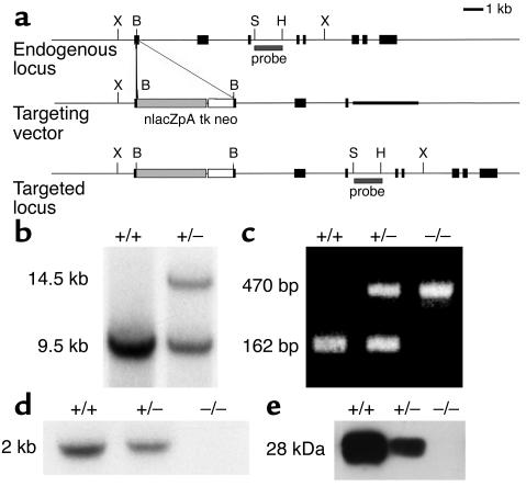
Figure 1.
Targeted disruption of the mouse MOG locus. (a) Partial restriction maps of the WT MOG allele (endogenous locus), the targeting vector, and the expected targeted locus. The location of the external screening probe is shown. X, XmnI; B, BstEII; S, SacI; H, HindIII. (b) Southern blot analysis of DNA from the parental ES cell line (+/+) and the targeted clone (+/–), digested with XmnI and hybridized with the external screening probe. This probe detects a WT 9.5-kb fragment or a 14.5-kb recombinant fragment. (c) Ethidium bromide–stained PCR products from tail DNA. The 470-bp and 162-bp fragments correspond to the mutated and the WT alleles, respectively. (d) Northern blot analysis of RNA isolated from the brain of MOG+/+, MOG+/–, and MOG–/– mice probed for MOG transcripts. (e) Western blot of brain proteins from 1-month-old MOG+/+, MOG+/–, and MOG–/– mice probed with anti-MOG antibodies.

