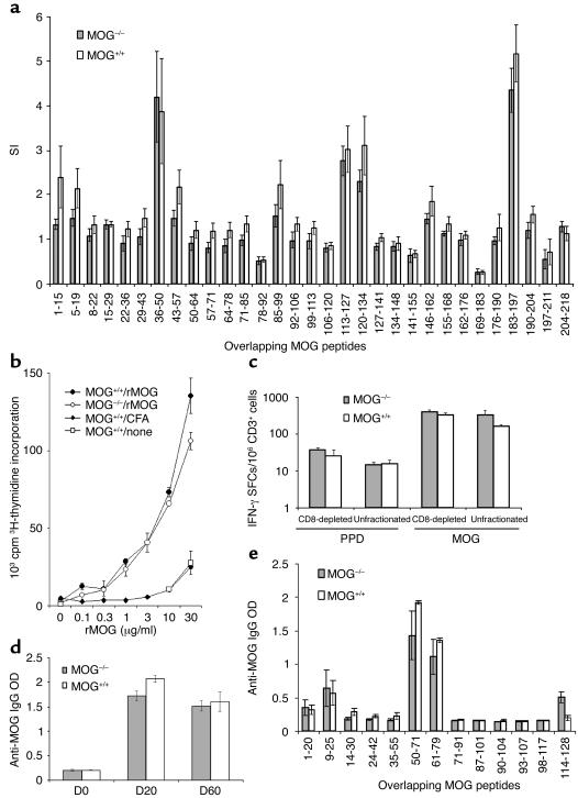
Figure 5.
(a) Proliferative response of lymph node cells MOG+/+ (n = 6) and MOG–/– (n = 6) mice 10 days after immunization with 31 overlapping mouse MOG peptides. The in vitro recall response was induced using individual MOG peptides (200 μg/ml). SI, stimulation index. (b) The proliferative response of lymph node cells of MOG+/+ (n = 3) and MOG–/– (n = 3) mice 10 days after immunization with mouse rMOG. The recall response was elicited with different doses of rMOG. (c) The MOG 35–55–specific T cell population was enumerated by IFN-γ ELISPOT in splenocyte cultures of individual MOG+/+ (n = 4) and MOG–/– (n = 3) mice 14 days after MOG 35–55 immunization. Unfractionated or CD8-depleted spleen cultures were stimulated with purified protein derivative (PPD) (50 μg/ml), or MOG 35–55 (50 μg/ml). (d) MOG-specific antibody response in MOG+/+ (n = 4) and MOG–/– (n = 4) mice at days 0, 20, and 60 after immunization with rat rMOG. MOG-specific IgG levels were assessed by ELISA on serum diluted 1:60; quantitatively similar results were obtained with 1:360 and 1:2,160 dilutions (data not shown). (e) Analysis of the fine specificity of anti-MOG IgG in MOG+/+ and MOG–/– mice 40 days after immunization with rat rMOG. Peptide-specific IgGs were assessed by ELISA with a panel of 13 overlapping rat MOG peptides. Comparable results were obtained at days 20 and 60 after immunization.

