Figure 7.
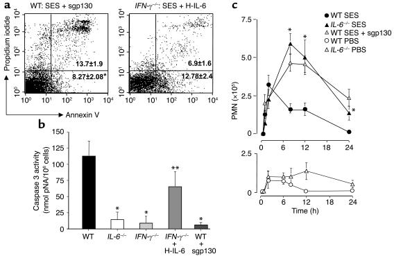
Modulation of IL-6 signaling in vivo affects apoptotic clearance of PMN in SES-induced peritoneal inflammation. (a) Annexin V/PI staining of leukocytes. Representative scatter plots show similar analysis performed on cells elicited from wild-type mice after 6 hours of stimulation with SES in combination with sgp130 (150 ng per mouse) or IFN-γ–/– mice after stimulation with SES in combination with HYPER-IL-6 (40 ng per mouse). (b) Caspase 3 activity was evaluated in isolated leukocytes from wild-type, IFN-γ–/–, and IL-6–/– mice intraperitoneally administered with SES, IFN-γ–/– mice administered with SES in combination with HYPER-IL-6 (40 ng per mouse), or wild-type mice administered with SES in combination with sgp130 (150 ng per mouse). Data (expressed as nanomoles of pNA per 106 cells) is presented from the 6-hour time point (mean ± SEM, n = 5 mice per condition; *P < 0.05, a significant decrease versus wild type plus SES; **P < 0.05, a significant decrease versus IFN-γ–/– plus SES). Inclusion of the caspase 3 inhibitor DEVD-fmk to wild-type leukocyte extracts reduced activity to background levels (data not shown). (c) Wild-type mice were intraperitoneally administered SES, SES plus sgp130 (150 ng per mouse), or PBS, and IL-6–/– mice were intraperitoneally administered SES or PBS. At defined intervals, PMN infiltration was assessed by differential cell count. The lower panel shows PMN influx in response to PBS alone. Results are expressed as the mean ± SEM of 12 mice per treatment (*P < 0.05 versus wild-type mice).

