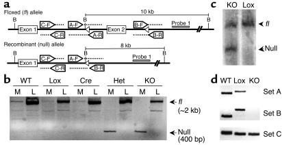
Figure 1.
Recombination of PPARγ-loxP allele in the muscle of MuPPARγKO mice. (a) Schematic of recombinant mouse PPARγ alleles indicating PCR primers sets, BamHI restriction sites (labeled as B), loxP sites (open triangles), and Probe 1 for Southern blot analysis. (b) Detection of intact (fl) versus recombined (null) allele by PCR on genomic DNA from muscle (M) or liver (L). The fl and null alleles appear at approximately 2 kb and 400 bp, respectively. (c) Southern blot analysis of BamHI-digested genomic DNA isolated from muscle samples from Lox and MuPPARγKO (KO) mice. Hybridization was performed with Probe 1, identifying fragments of 10 and 8 kb for fl and null alleles, respectively. (d) PCR detection of unrecombined alleles (primer sets A and B) or any WT, fl, or null allele (primer set C) in genomic DNA isolated from enriched myocytes of WT, Lox, and KO mice. Primer sets A and B yield larger products in the fl compared with WT allele due to loxP insertion.

