Figure 5.
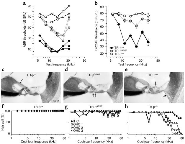
Cochlear function and histopathology in TR-β mutant animals. (a) ABR thresholds for TR-βGS/GS and TR-β–/– mice are elevated with respect to TR-β+/+, but loss is greater in TR-β–/–. Data from individual 8-week-old animals are shown. (b) DPOAE thresholds for TR-βGS/GS and TR-β–/– mice are elevated with respect to WT; however, TR-β–/– exhibited a much greater deficit. Data are mean ± SEM. Group sizes were 10, 18, and 14 for TR-β+/+, TR-βGS/GS, and TR-β–/–, respectively. (c–e) Photomicrographs of the upper basal turn (cochlear frequency approximately 16 kHz) from WT and TR-β mutant cochleas. Arrows indicate: (c) tectorial membrane in TR-β+/+; (d) misaligned feet of outer pillar cells in TR-βGS/GS; (e) collapse of outer supporting cells (unfilled) and loss of spiral ligament fibrocytes in TR-β–/– (filled). Scale bar in c applies to all three images. (f–h) Basal-turn OHC loss is seen in TR-β–/– mice. Data from one ear of each genotype are shown. Symbol key in g applies to all three panels.

