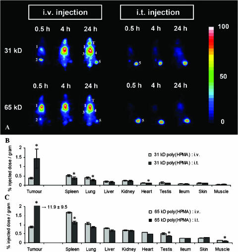
Figure 2.
Effect of i.t. injection on the biodistribution of HPMA copolymers. (A) Scintigraphic analysis of the effect of i.t. injection on the biodistribution of 31-kDa and 65-kDa poly(HPMA) in rats bearing subcutaneously transplanted AT1 tumors. In the images obtained 0.5 hour after i.v. administration, the accumulation of the radiolabeled copolymers was most prominent in the heart (i.e., circulation) (1) and bladder (2). In the images obtained at 4 and 24 hours, the highest amounts of the copolymers were found in the heart/lungs (1), spleen (3), liver (4), and tumor (5). In addition, at the two latter time points, released radioactive iodine was found to accumulate in the thyroid (T). On i.t. injection, only localization to the tumor (5) could be observed over the first 24 hours after administration. (B and C) Quantification of the effect of i.t. injection on the tumor and organ concentrations of 31-kDa poly(HPMA) (B) and 65 kDa-poly(HPMA) (C) at 24 hours p.i. Values represent the average ± SD of four to six animals per experimental group. *P < .05 vs i.v. injection (Student's t test).

