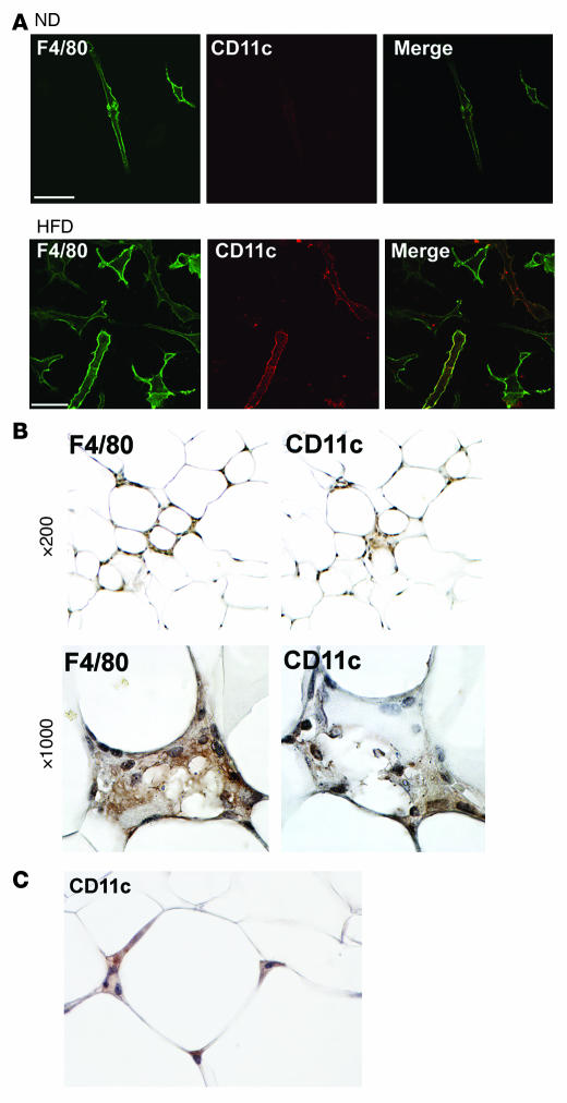
Figure 2. CD11c+ ATMs in SVF cultures and in adipose tissue from obese mice.
(A) Identification of F4/80+ and CD11c+ ATMs by immunofluorescence microscopy. Epididymal fat pads from ND and HFD mice were separated into adipocyte and SVF fractions. SVF cells were plated onto glass coverslips and cultured overnight prior to fixation. Cells were stained with antibodies against F4/80 (left) and CD11c (middle) and imaged by confocal microscopy to identify surface markers to confirm the presence of CD11c+ cells only in the SVF from HFD-fed animals. Similar results were obtained for 3 independent sets of cultures. (B) Immunohistochemical localization of CD11c+ in adipose tissue. Consecutive sections from epididymal fat pads from obese C57BL/6 mice were stained with anti-F4/80 (left panels) and anti-CD11c antibodies (right panels), followed by colorimetric detection (brown). Sections were counterstained with hematoxylin (blue) and images taken at low (×200) and high magnification (×1,000). (C) In obese mice, CD11c+ cells were also detected surrounding normal-appearing adipocytes in the absence of crownlike macrophage clusters (×1,000 magnification).

