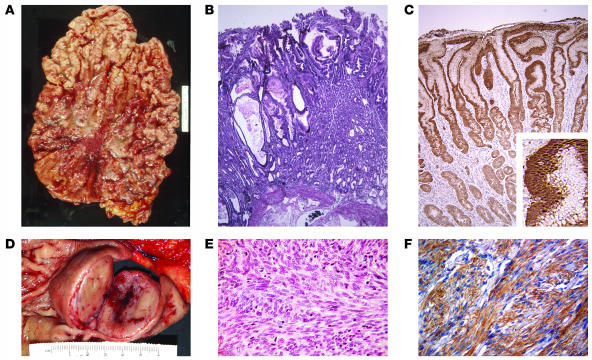
Figure 2. Gross and microscopic view of Ménétrier disease and GIST.
(A and B) Gross (A) and microscopic (B) appearance of the stomach of a patient with Ménétrier disease (original magnification of B, ×40). (D and E) Gross (D) and microscopic (E) appearance of the stomach of a patient with a submucosal gastric GIST (original magnification of E, ×200). In D, the mucosa is stretched over the submucosal GIST with its typical central degenerative changes. (E) Typical spindle-shaped appearance of a GIST; less common are epithelioid-shaped and mixed epithelioid- and spindle-shaped GISTs. (C) TGF-α immunoreactivity in the gastric mucosa of a Ménétrier disease patient (original magnification, ×100; inset, ×300). (F) KIT immunoreactivity in a GIST (original magnification, ×200). The rulers in A and D are the same scale.

