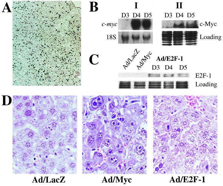
Figure 1.
(A) β-Galactosidase staining of day 5 Ad/LacZ liver. (B) (I) Northern blot showing exogenous MYC gene expression starting on day 4. 18S is shown as loading control. (II) Western blot showing c-Myc protein expression in Ad/Myc livers. Coomassie staining is shown for loading control. D3, D4, and D5 indicate total RNA and protein samples isolated from Ad/Myc livers killed on days 3, 4, and 5, respectively. (C) Western blot showing that E2F-1 is expressed by day 3 postinjection of recombinant adenovirus carrying E2F-1. E2F-1 is not detectable in Ad/LacZ or Ad/Myc livers. (D) Hematoxylin/eosin staining of day 4 Ad/LacZ, Ad/Myc, and Ad/E2F-1 liver. (Magnification: A, ×5; D, ×40.)

