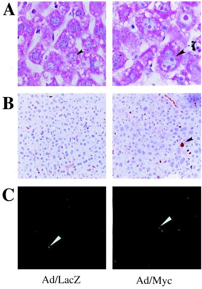
Figure 2.
(Left) Day 4 Ad/LacZ liver. (Right) Day 4 Ad/Myc liver. (A) Methyl green pyronin staining of the sections. The arrows highlight enlarged nucleoli of Ad/Myc hepatocytes. (B) Ki-67 staining of liver sections. The arrow indicates positive staining of a proliferating cell. (C) TUNEL staining of liver sections. The arrows indicate nuclei of cells undergoing apoptosis. (Magnification: A, ×60; B and C, ×10.)

