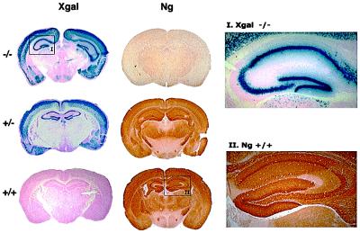
Figure 2.
Staining of the mouse brain sections with X-gal and Ng-specific antibody. Coronal sections through the hippocampus of the WT (+/+), HET (+/−), and KO (−/−) mice were stained with X-gal or with Ng-specific antibody (no. 2641). The magnified areas show a good correspondence between the X-gal and Ng-positive staining of neurons between +/+ and −/− mice. X-gal staining is restricted to neuronal cell bodies, whereas Ng-positive staining covers both cell bodies and processes.

