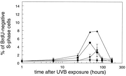
Figure 3.
Mice were exposed to 250 J/m2 UVB and epidermal cells were isolated at various times after UVB exposure. One hour before cell isolation, mice received a single dose of BrdUrd to identify S-phase cells, and the percentage of BrdUrd-negative S-phase cells from WT (●), Xpa−/− (▴), Xpc−/− (▾), and Csb−/− (■) mice was determined. Data are means ± SEM (n ≥ 3) or variation (n = 2).

