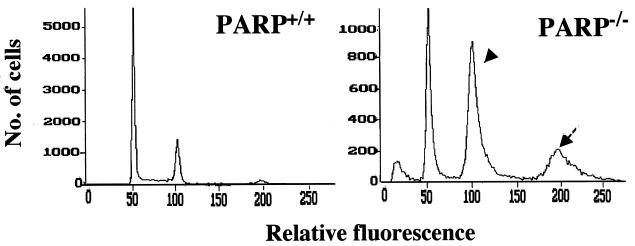
Figure 1.
Flow cytometric analysis of primary wild-type and PARP−/− fibroblasts. Asynchronously dividing cells were grown to ≈60% confluency for 3 days, after which nuclei were prepared and stained with propidium iodide for flow cytometric analysis. In addition to the major peak corresponding to G0-G1 (haploid) nuclei apparent for wild-type cells, PARP−/− cells exhibited a larger peak corresponding to G2-M (diploid) nuclei (arrowhead) as well as a third peak corresponding to tetraploid nuclei (arrow).

