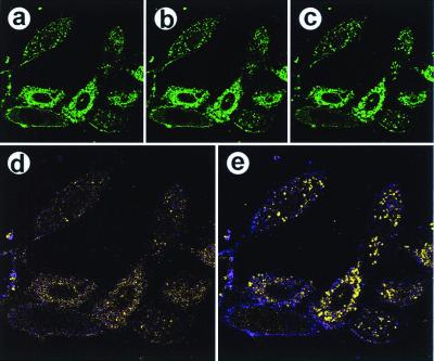
Figure 3.
PDGF stimulation of NIH 3T3 fibroblasts causes a relocalization of PtdIns(4,5)P2-NBD. Serum-starved (3 h) NIH 3T3 cells were treated with a complex of PtdIns(4,5)P2-NBD (10 μM) and histone (3 μM). After 10 min, cells were stimulated with 380 ng/ml PDGF; the medium was not changed subsequently. (a) An image before PDGF addition (t = 0), using the 488-nm laser line. The same optical section (Z-section) was collected for 20 min at 10-sec intervals. b and c were obtained at t = 3 min and 15 min, respectively. (d) A difference image calculated by using the t = 0 and t = 3-min data, in which fluorescence intensity corresponds to movement of the PtdIns(4,5)P2 or its metabolic products. Thus, purple represents a decrease in fluorescence and yellow represents an increase in fluorescence in the 3-min image relative to the zero-time image. (e) A difference image comparing the 15-min data with the zero-time data. Magnifications: a–c, ×240; and d and e, ×360.

