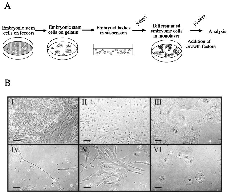
Figure 1.
Induced differentiation of human ES-derived cells in culture. (A) A schematic representation of the differentiation protocol (see Materials and Methods). (B) Various morphologies of ES cells before and after induced differentiation: I, a colony of ES cells on feeders; II–VI, differentiated cells cultured in the presence of HGF, activin-A, RA, bFGF, or BMP-4, respectively. Note the small size of ES cells in the colony in comparison with the size of the differentiated cells. Scale bar = 100 μm.

