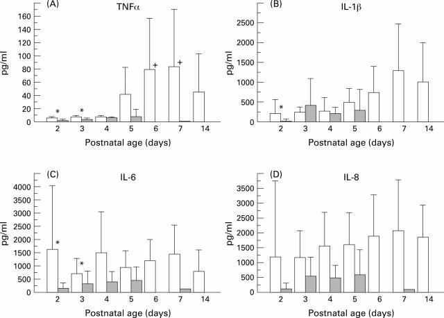
Figure 1 .
TAF fluid concentrations of proinflammatory cytokines in infants who subsequently developed CLD and infants with uncomplicated RDS (shaded area). Bars indicate mean values with 95% confidence intervals, on separate days. (A) TNFα concentrations: *denotes p<0.05 on days 2 and 3 between CLD and RDS infants; + p>0.05 on days 6 and 7 compared with days 2 and 3 for CLD infants. (B) IL-1β antigen titres:*p <0.05 between RDS and CLD infants on day 2. (C) IL 6 concentrations: *p<0.05 on days 2 and 3 between the two groups of infants. (D) IL 8 concentrations

