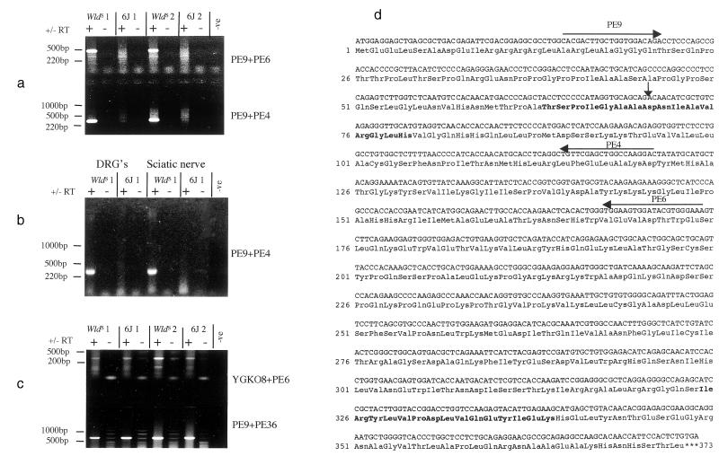
Figure 2.
(a) RT-PCR showing the detection in brain of a WldS-specific chimeric transcript between Ufd2 and D4Cole1e. Primers indicated in (d) were used to amplify fragments of 478 bp and 318 bp as shown. Samples derived from different animals are distinguished by the labels WldS 1, WldS 2, etc. (b) RT-PCR showing WldS-specific expression of the chimeric transcript in dorsal root ganglia (DRG) and sciatic nerve. (c) RT-PCR showing the expression of normal transcripts in both 6J and WldS brain for D4Cole1e (Upper) and Ufd2 (Lower) (expected sizes 334 and 710 bp, respectively). Primer sequences: YGK08 = 5′-ACTAGGGCCGTTTGGCTTC-3′; PE36 = 5′-CCTGAACTGGAGCCAGTGTT-3′; PE6 and PE9 as shown in (d). (d) Sequence of the chimeric cDNA and the predicted coding sequence. The vertical arrow indicates the junction between Ufd2-derived sequence and D4Cole1e-derived sequence (Asp-71 is derived from neither protein but formed by the junction). Horizontal arrows indicate the location, and 3′ direction of primers used in (a) and amino acids shown in bold are those used to make peptides for generation of polyclonal antisera 183 (Ile-325 to Lys-339) and 185 (Thr-64 to His-79).

