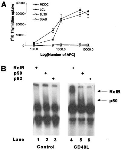
Figure 1.
APC function and RelB location in BL cell lines. (A) Varying numbers of either MDDC, EBV-transformed lymphocytes (LCL), or the BL cell lines BJAB or BL30 were incubated with 105 purified allogeneic T cells for 5 days, and T cell proliferation was measured by incorporation of [3H]thymidine. Data are the mean of triplicate wells ± SD and are representative of three separate experiments. (B) Nuclear extracts were prepared from BJAB after incubation for 24 h in the presence or absence of 50 ng/ml sCD40L. For supershift assays, 10 μg of nuclear extract was incubated with either 2 μg of RelB, p50, or p52 antibody for 1 h at 4°C, followed by labeled NF-κB probe, and separated on a 4% polyacrylamide gel. No supershift was demonstrable in the presence of rabbit Ig control (not shown).

