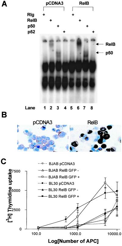
Figure 2.
Overexpression of RelB enhances APC function of BL cell lines. (A) BJAB were transfected with either RelB or pCDNA3 and incubated for 24 h before preparation of nuclear extracts, incubation with antibody and labeled NF-κB probe, and separation as in Fig. 1B. (B) BJAB were transfected with either RelB or pCDNA3, together with the GFP-expressing plasmid pGreenLantern, and incubated for 24 h; then, GFP+ cells were sorted by flow cytometery. An amount equal to 105 of each population of GFP+ cells was cytospun, fixed, and stained for RelB by using an immunoperoxidase technique. RelB is stained with diaminobenzidine (brown), and the nucleus is counterstained with hematoxylin (blue) at magnification ×130. (C). BJAB and BL30 cell lines were transfected, and GFP+ and GFP− cells were sorted and incubated with allogeneic T cells as in Fig. 1. Data are the mean of triplicate wells ± SD and are representative of two separate experiments.

