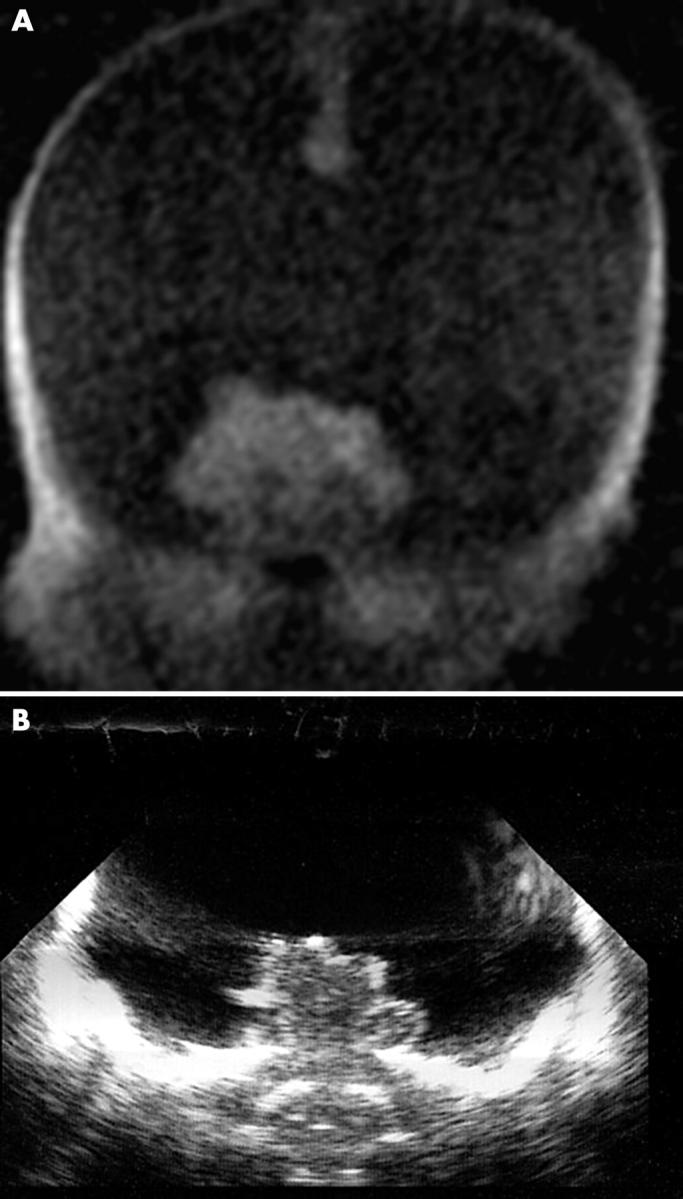
Figure 5 .

T1 weighted coronal image (A) and ultrasound image (B). Hydranencephaly is seen on both imaging modalities, but was easily detected by magnetic resonance. Massive hydrocephalus could not be completely excluded by ultrasound (although it was less likely).
