Figure 1.
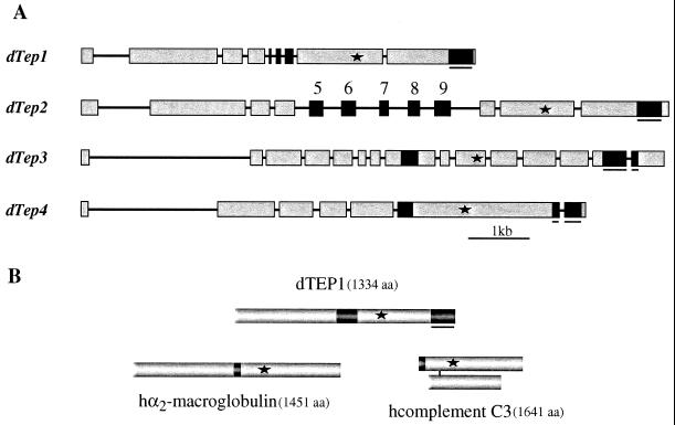
(A) Gene organization of Drosophila Tep1, -2, -3, and -4. Exons are represented by boxes and introns by lines. Asterisks indicate the positions of the thiolester motifs. Black nonunderlined boxes correspond to the variable region of TEPs. Five isoforms of Tep2 transcripts have been isolated; each transcript differs only by a single exon (exons 5 to 9) (E.P. and M.L., unpublished data). Black underlined boxes correspond to the highly conserved C-terminal region containing six cysteines. Chromosome localizations are as follows: Tep1, 35F1-F4; Tep2 and 3, 28B1-B4 (these genes are positioned at 2 kb from one another in reverse orientation); Tep4, 37F1-F2. (B) Protein structures of the three subfamilies of thiolester-containing proteins represented by Drosophila TEP1, human α2-macroglobulin, and human complement C3. The asterisks indicate the positions of the thiolester motifs, the black boxes show the respective positions of the variable domain in TEP, the bait domain of α2-macroglobulin, and the anaphylatoxin of complement C3. The underlined black box corresponds to the particularly well conserved region in TEPs (see Fig. 2).

