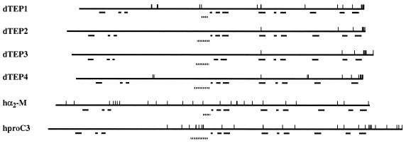
Figure 3.
Schematic representation of the protein structure of Drosophila TEP1 to -4, human α2-macroglobulin, and the proprotein for human complement C3; vertical bars show the positions of cysteine residues; horizontal bars indicate the positions of the various domain-signatures defined for the superfamily of thiolester-containing proteins. From left to right, blocks A to L (see also file 1, supplementary Fig. 6). Dotted lines show the positions of hypervariable regions in TEPs, the position of the bait domain of α2-macroglobulin, and anaphylatoxin in complement C3.

