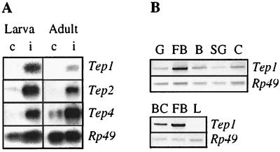
Figure 4.
(A) Transcription levels of Drosophila Tep1, -2, and -4 before (c) and after (i) an immune challenge (6 h) in wandering larvae, and in 6-day-old adult wild-type Drosophila. Five micrograms of polyA-RNA were fractionated by electrophoresis in denaturating agarose-formaldehyde gels. After transfer to a nylon membrane, the RNAs were hybridized with random-primed [32P]cDNA probes corresponding to Tep1, -2, and -4; and Rp49 for the loading control. (B) Transcription level of Tep1 in different tissue extracts of immune-challenged larvae: G, gut; F, fat body; B, brain; SG, salivary glands; C, carcass; BC, blood cells; and L, l(2)mbn cells challenged with bacteria. DNA fragments were amplified by PCR with specific primers of Tep1 and separated on agarose gel. The Rp49 amplification product served as an internal control.

