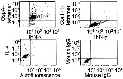
Figure 5.
Tetramer staining combined with intracellular cytokine staining on a representative OspA-reactive T cell clone. T cell clone was activated in vitro for 48 h; at 36 h, monensin was added; then during the last 2 h, 20 μg/ml OspA or control (LFA-1) tetramer was added. Cells were fixed, stained with directly labeled antibodies specific for IFN-γ, IL-4, and isotype controls for 30 min at 4°C, and washed twice before fluorescence-activated cell sorter (FACS) analysis. This clone did not respond to LFA-1 in in vitro proliferation assays.

